


 0752-2228195
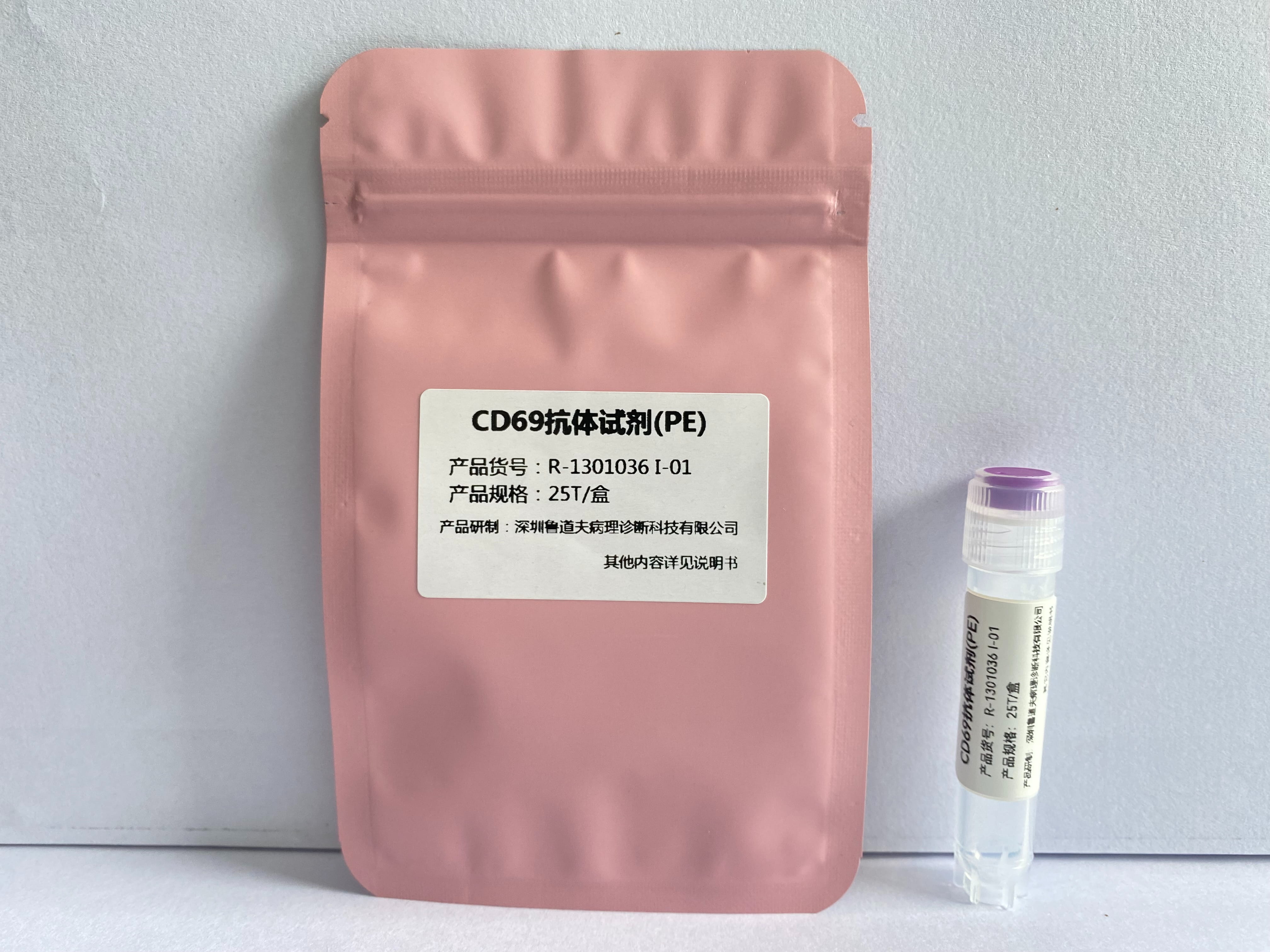
CD69 antibody reagent (PE)
Wholesale Price(RMB):
5161132
Product Code:R-1301036Ⅰ-01/R-1301036Ⅰ-02
File Number:

5161132
| Product Name | Anti-Human CD69, PE Flow Cytometry Antibody |
|---|---|
| Target | CD69, CLEC2C |
| Label | PE |
| Instrument | Suitable for flow cytometers with 488nm, 532nm, 561nm lasers |
| Subtype | Mouse IgG1, κ |
| Clone Number | FN50 |
| Recommended Application | Flow Cytometry |
| Dosage | 5 μL /Test |
| Purification | Affinity Chromatography |
| Stock Solution | Phosphate buffer solution containing 0.2% BSA and 0.2% proclin 950 (pH7.2) |
| Storage Conditions | Store at 2 - 8℃, protect from light, do not freeze |
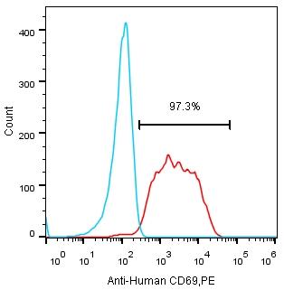
Fig.-After the anticoagulant of human heparin was stimulated by PMA/Ionomycin, the control group of the same type was stained with Mouse IgG1 Isotype Control, PE (left peak) and the experimental group with Anti-Human CD69, PE (Clone FN50) (right peak).
Overview of CD69 molecular target information
Molecular name: CD69, CD69 molecule
Gene family: CD molecules; C-type lectin domain containing
Alias: CLEC2C
Full name: CD69 antigen (P60, early t-cell activation antigen)
Review of CD69 molecular targets
CD69 is a type II transmembrane glycoprotein, belonging to the family of C- lectin receptors and a member of the complex family of NK cell signal transduction genes. It is the earliest surface antigen expressed by T lymphocytes after activation. When expressed, it can be used as a co-stimulatory signal to promote the further activation and proliferation of T lymphocytes. CD69 was also induced to express on NK cells, B cells, macrophages, neutrophils and eosinophils. The activated T cells expressing CD69 are mainly CD8+CD 45 RO+,and are mainly induced by IL-15 and related to the contact with activated endothelium expressing ICAM-1. When the exogenous stimulus disappears, the expression of CD69 in activated T cells can decrease rapidly. Therefore, the degree of CD69 expression in lymphocytes after exposure to an antigen can reflect the degree of cell response to the antigen.


Copyright: Shenzhen Rudolf Pathological Diagnosis Technology Co., Ltd


