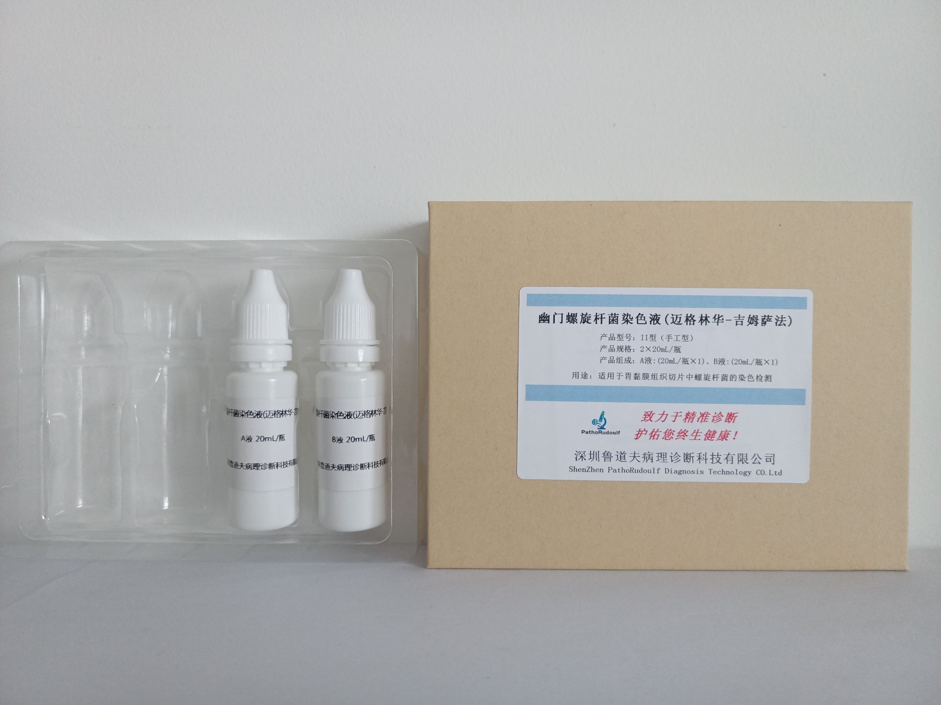
Gastric Helicobacter Pyloric (Helicobacter Pyloric, HP), also known as gastric Campylobacter Pyloric (Campylobacter Pyloric), it has been proved that this bacterium and chronic gastritis and peptic ulcer have a close relationship, gastric Helicobacter Pyloric is generally arc-shaped, S-shaped or seagull-shaped, and sometimes 3 to 4 curved helical, often fish school-like distribution. The bacterium is mostly found in the gastric mucosa between the surface epithelium and the mucosal layer, and close to the surface epithelial cells, part of the epithelial cells into the cytoplasm, the gastric concavity and the mucous membrane in the superficial gland lumen of the bacterium. Helicobacter pylori staining is mainly methylene blue method, silver nitrate method, May-Grunwald-Giemsa method (May-Grunwald-Giemsa, MGG method), alkaline magenta method, etc. The silver nitrate method is clear, the stained film can be preserved for a long time, but the operation is more cumbersome and time-consuming. Other methods are easier, but the stained film is easy to fade.
The staining solution is mainly composed of Maiglinhua stain and Giemsa stain, after staining gastric H. pylori is blue, collagen fibers are red, red blood cells are green, gastric mucosal epithelium is light blue, the nucleus of the cells is dark blue, HP is mostly located in the mucus on the surface of the gastric mucosal epithelium, especially in the gastric microcavity of the number of more.


 0752-2228195