Sporozoites have no obvious motile organelles and live parasitically throughout their developmental stages. The life history is complex, with asexual proliferation of schizonts, spore proliferation and sexual gamete reproduction, and can be accomplished in the same host or in two different hosts. The main parasitic sporozoites are Plasmodium, Toxoplasma gondii, Pneumocystis carinii, Cryptosporidium and so on.
Plasmodium is a parasitic sporozoite, mainly parasitizing humans and many kinds of mammals, with a few parasitizing birds and reptiles, of which more than 130 species are known. There are more than 130 known species of Plasmodium parasites in humans, including Plasmodium vivax, Plasmodium falciparum, Plasmodium vivax and Plasmodium ovale. The basic structure of Plasmodium consists of nucleus, cytoplasm and cytosol. After staining with Riesling or Giemsa, the nucleus is purplish red and the cytoplasm is blue. Plasmodium develops successively in hepatocytes and erythrocytes in the human body, and its pathogenicity occurs during the proliferation of intraerythrocytic stage schizonts. The development of Plasmodium in erythrocytes consists of four stages: microtrophozoites, macrotrophozoites, schizonts and gametocytes, which can be observed by Riesling or Giemsa staining of different morphologies in different periods.
The pathogenetic examination of Plasmodium is mainly divided into thick and thin blood film staining microscopy method and quantitative analysis of blood sedimentation brown layer (QBC). Thick blood film in the protozoa is more concentrated, easy to detect, but the preparation of erythrocytes are easy to dissolve, the morphology of the protozoa changed, species identification is more difficult; thin blood film in the erythrocytes are not easy to be destroyed, the morphology of Plasmodium protozoa structure is more complete, typical, easy to recognize and identify the species, but the protozoa density of the low is easy to miss the detection; therefore, it is often used to make a slide at the same time thick, thin two kinds of blood film, with Ruichter's, Giemsa or acridine orange staining to find the presence of Plasmodium. The principle of QBC analysis is that the red blood cells infected with Plasmodium are lighter than normal red blood cells and slightly heavier than white blood cells, after centrifugation and stratification, they are concentrated in the upper part of the normal red blood cell layer and the lower part of the white blood cell layer, and the results are observed by fluorescence microscope after adding acridine orange reagent, which is a lot more sensitive than the common microscopic method and is more simple and quicker, but with a higher cost.
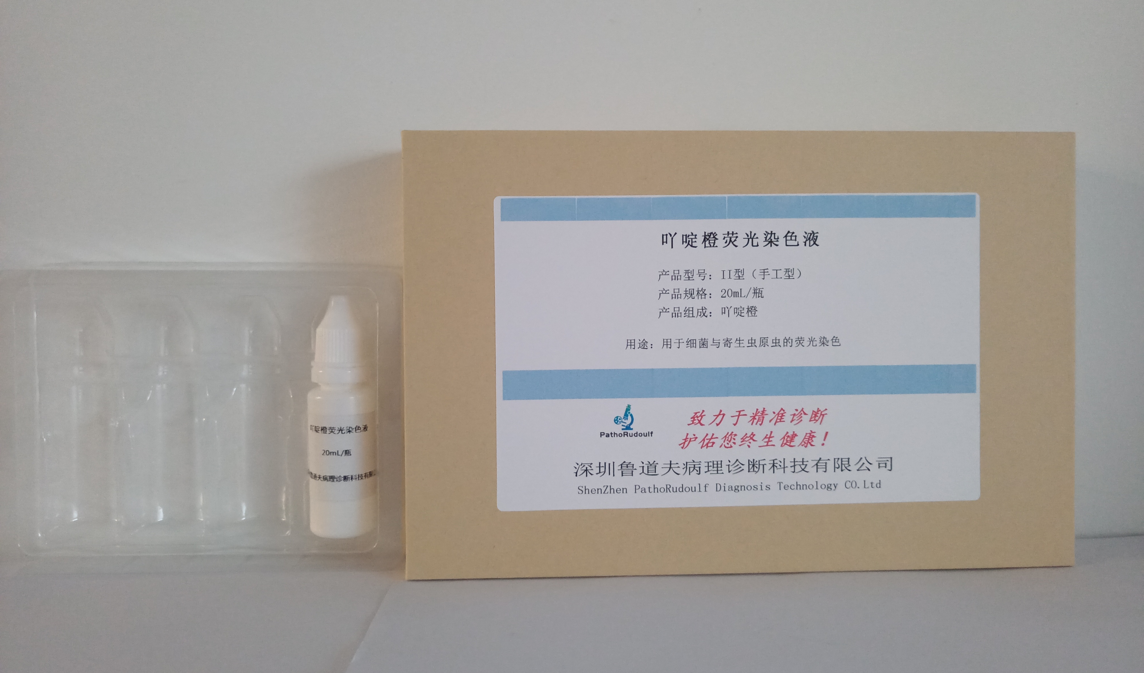
Plasmodium staining solution (acridine orange staining solution) consists of acridine orange and phosphate, etc. It mainly adopts the fluorescent dye acridine orange for staining the thick and thin blood membrane, which combines with different components of parasites or tissue cells through osmosis, adsorption and chemosynthesis to become a fluorescent specimen, and then through the excitation of fluorescent light source, the specimen of the combined fluorescein emits fluorescence, and then the presence of Plasmodium can be observed by fluorescence microscope in the dark room. The presence of Plasmodium can be observed through fluorescence microscope in the dark room. This method is simple and quick, and the detection rate is higher than the conventional method.


 0752-2228195