Muscle fiber (Muscle fiber) belongs to the muscle tissue components, composed of myoblasts, according to the morphological and functional characteristics, muscle fibers can be divided into smooth muscle (also known as transverse muscle), skeletal muscle, cardiac muscle; myofiber staining methods have a variety of methods, such as Li Chun red, aniline blue, tungsten phosphorotungstic acid hematoxylin method, etc., tannins - azofluorescent peach method is the use of the two acid dyes successive role in the completion of the identification of the staining. The method of tannin-azo-fluorescent peach is to use two acidic dyes to complete the identification of staining.
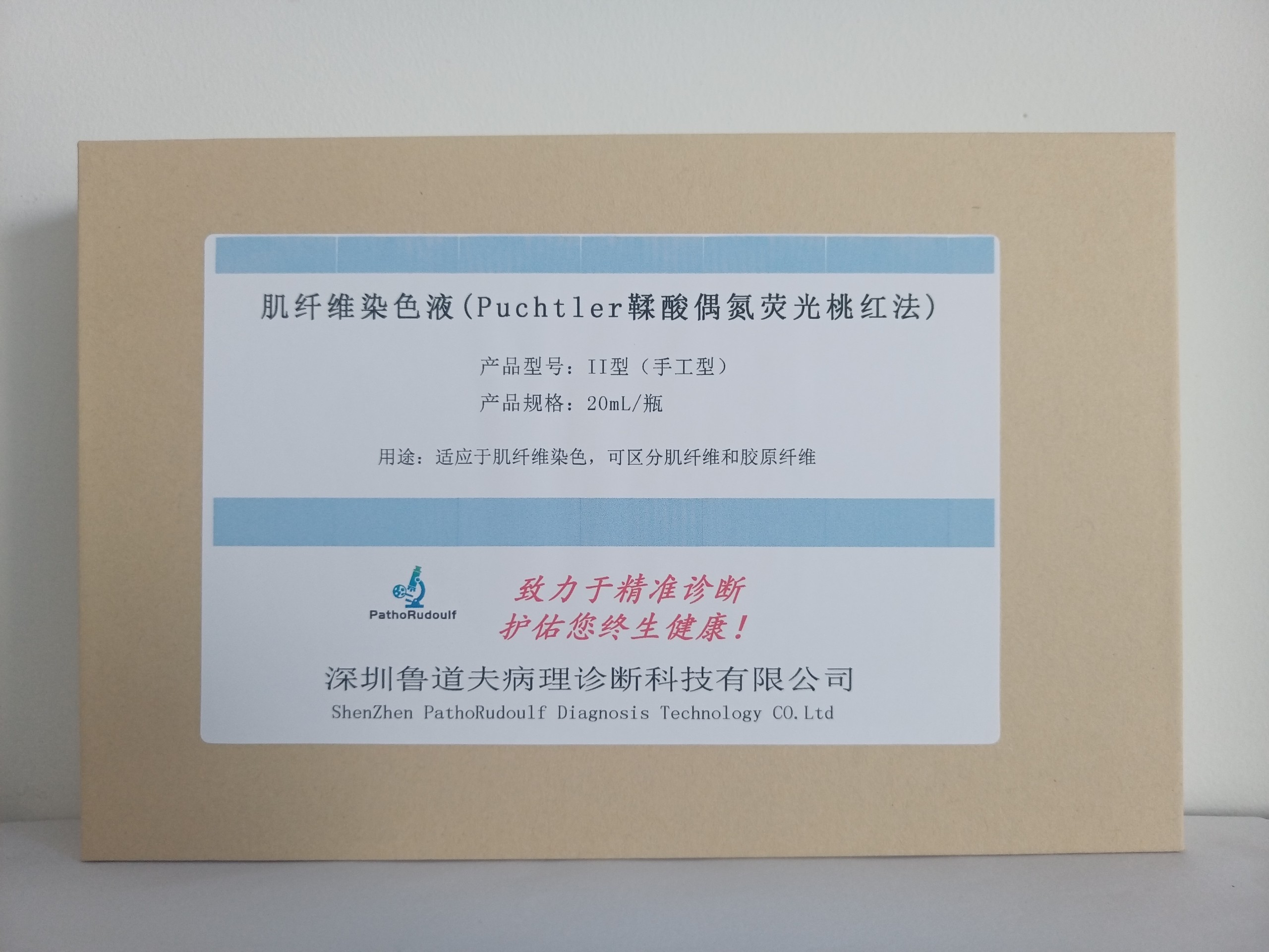
Myofibrillar staining solution (Puchtler ellagic acid azo-fluorescent peach method) is mainly composed of Lea hematoxylin staining solution, ellagic acid differentiation solution, phosphomolybdic acid differentiation solution, azo-fluorescent peach staining solution, etc. Its staining principle lies in the fact that ellagic acid is easy to enter into the permeability of the collagen fibers, and collagen fibers are yellow, while the azo-fluorescent peach stain is easy to enter into the permeability of the muscle fibers is lower, and muscle fibers are red. The staining solution is used to distinguish myofibers and collagen fibers, the contrast is clear, should not fade, and at the same time can show the myoepithelial cells, which can be used for the diagnosis of myoepithelial tumors of the breast and skin.


 0752-2228195