Mast cells are common cells in loose connective tissues, often distributed in groups along small blood vessels and lymphatic vessels, and are also commonly found in bronchial tubes and around the interlobular ducts of the pancreas. Generally, the cells are large, with diameters of about 20-30 μm, round or oval, with small nuclei, and cytoplasmic cytoplasm filled with coarse and heterochromatic eosinophilic particles.
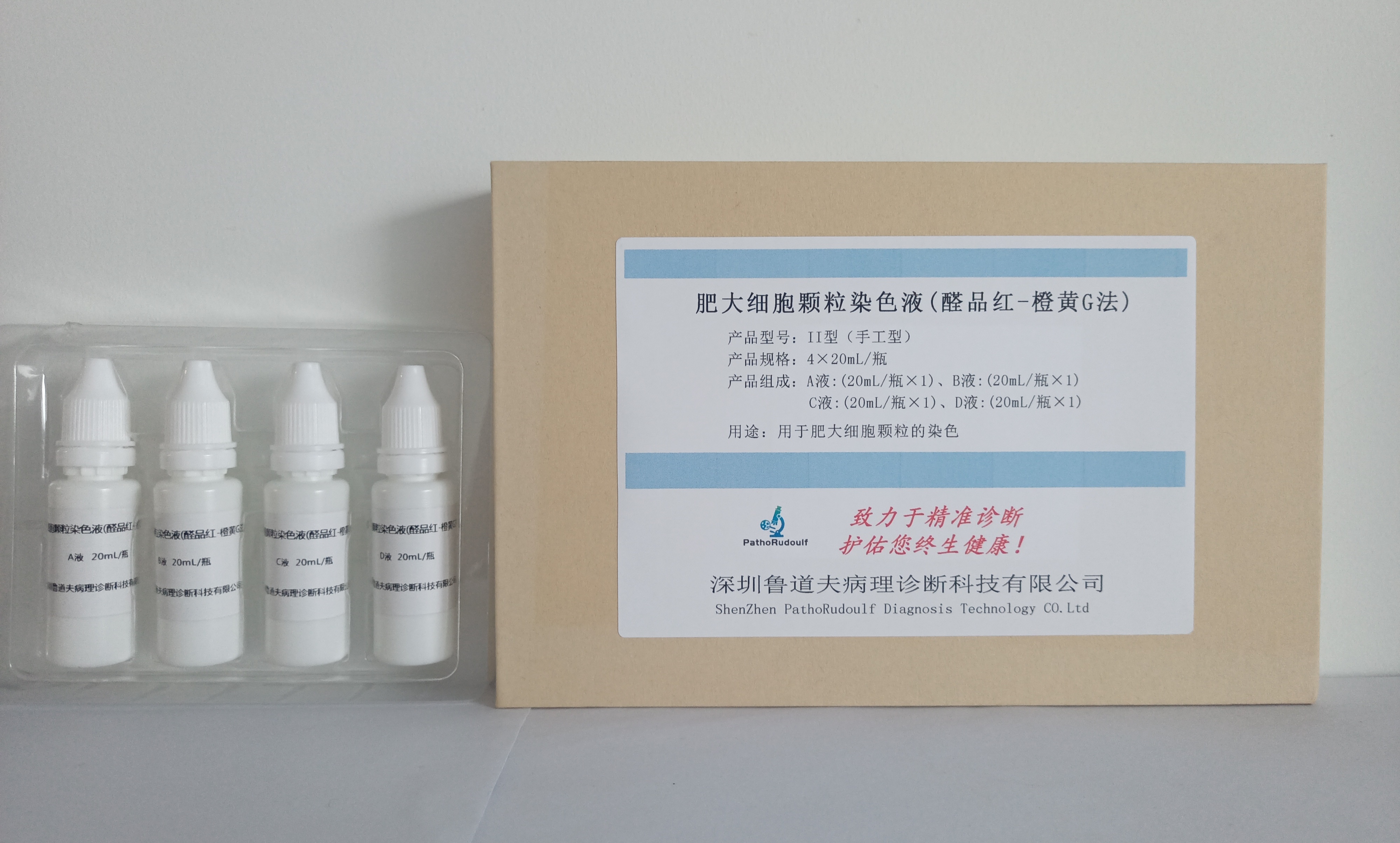
The staining mechanism of mast cell staining solution (aldehyde magenta-orange yellow G method) lies in the fact that aldehyde magenta has a strong affinity for acidic mucopolysaccharides containing sulfate groups, and mast cell particles contain heparin with carboxyl and sulfate groups, so it is easy to combine with aldehyde magenta to form a complex and color; orange yellow G is an acidic dye, which can stain other tissue components into yellow. This method can show heterochromatic particles, after staining the mast cell granules are stained dark purple, other cells are not colored, the background is orange-yellow, contrast is more distinct.


 0752-2228195