Other solid tumors
AGTRAP-BRAF and SND1-BRAF fusion genes are present in gastric cancer and are associated with treatment with RAF/MEK inhibitors.
Colorectal cancer
In addition to BRAF fusions with KIAA1549 in central nervous system tumors, BRAF translocations are also found in colorectal cancer. Furthermore, BRAF gene translocations have been identified in some cases of Myxoinflammatory fibroblastic sarcoma (MIFS) lacking TGFBR3-MGEA5 translocations.
Central nervous system tumors
A molecular subtype of central nervous system gliomas, this fusion is observed in: pilocytic astrocytoma (common), pilocytic mucinous astrocytoma (sellar region), and diffuse ependymoma.
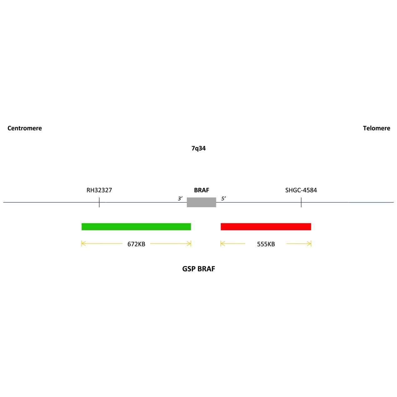
BRAF is an oncogene located at 7q34. Approximately 60%-80% of patients with pilocytic astrocytoma have been found to have KIAA1549-BRAF gene fusions. Detection of this fusion gene via FISH can play a role in the differential diagnosis of low-grade gliomas, as outlined in the “Chinese Guidelines for the Diagnosis and Treatment of Central Nervous System Gliomas (2015).” The KIAA1549-BRAF fusion is an important diagnostic marker. Since pilocytic astrocytomas also exhibit microvascular proliferation, they are difficult to distinguish from GBM histologically. If the KIAA1549-BRAF fusion is detected, it strongly suggests a pilocytic astrocytoma;
Note: Since BRAF and its partner gene KIAA1549 are only 2 M apart in the genome, it is difficult to distinguish between negative and positive cells under FISH microscopy using BRAF breakage probes. PCR detection is recommended. When BRAF fuses with other partner genes, it exhibits a typical 1 red, 1 green, and 1 yellow signal pattern.


 0752-2228195