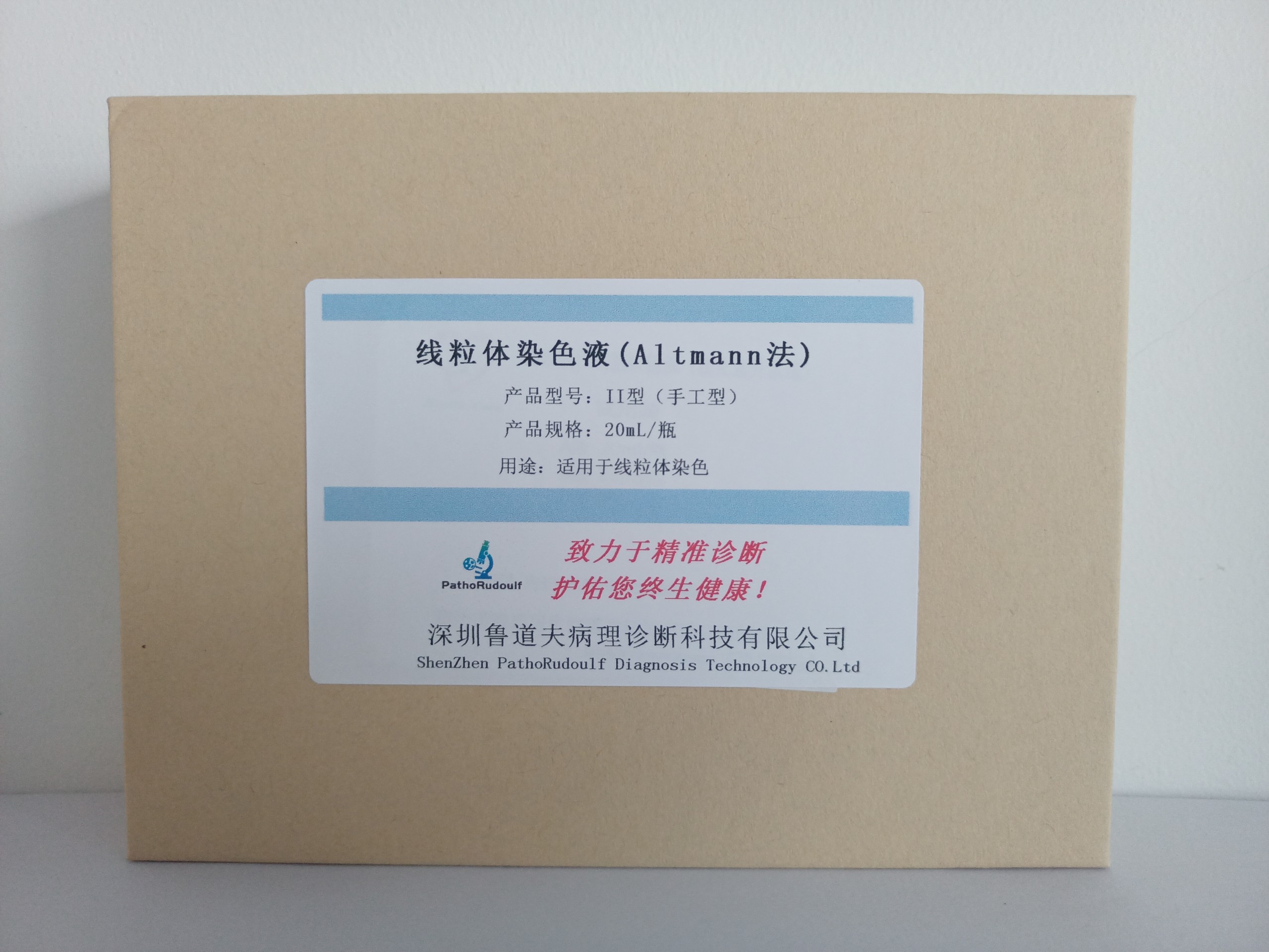
5609902300249560
3×50mL/bottle(Ⅰ-01)3×100mL/bottle(Ⅰ-02)3×250mL/bottle(Ⅰ-03)3×20mL/bottle(Ⅱ-01)3×50mL/bottle(Ⅱ-02)

5609902300249560
3×50mL/bottle(Ⅰ-01)3×100mL/bottle(Ⅰ-02)3×250mL/bottle(Ⅰ-03)3×20mL/bottle(Ⅱ-01)3×50mL/bottle(Ⅱ-02)
Mitochondria are the source of cellular energy and can vary greatly in shape (from rod-shaped to round). Mitochondria are very small and can only be visualized with the aid of an electron microscope.Mitochondria contain hereditary maternal DNA and vary in number, size and shape depending on the animal cell type. Mitochondria are best viewed with an electron microscope, and histopathologic methods such as the Altmann technique are helpful. Successful display of mitochondria by histochemical methods depends on several factors: the tissue must be freshly fixed, the sections are thin (2-3 μm), and since mitochondria are one of the earliest organelles to undergo degeneration when cells are hypoxic or die, rapid fixation is essential.
Mitochondrial staining is best achieved by the Champy-Kull method, but the staining technique is more complex.